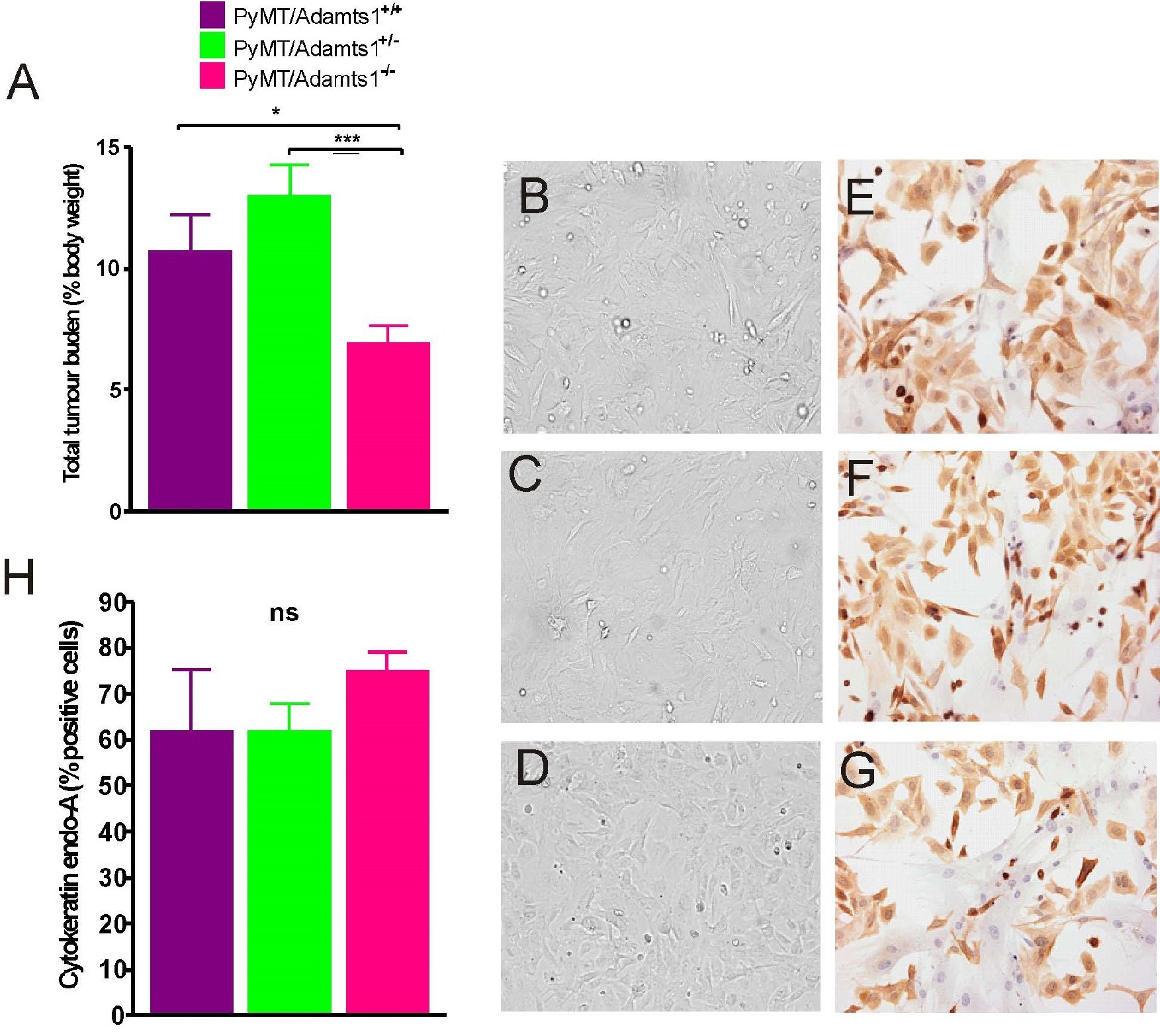
Fig. 3. PyMT/Adamts1-/- mice have smaller mammary tumors than Adamts1+/+ and Adamts1+/- littermates, and isolated 1omMCC were predominantly of mammary epithelial cancer cell type. a) Mammary tumors were excised from any of the 10 mammary glands where tumor growth was found and digested to isolate mammary epithelial cancer cells. Total tumor weight of Adamts1+/+ (n=10), Adamts1+/- (n=13) and Adamts1-/- (n=15) mammary tumors in proportion to body weight. b, c, d) Brightfield images of Adamts1+/+, Adamts1+/- and Adamts1-/- 1omMCC, revived from liquid nitrogen and grown under in vitro cell culture conditions. e, f, g) 1omMCC immunocytostained against epithelial cell marker, cytokeratin endo-A. Images were taken at 20x magnification. h) Proportion of mammary epithelial cancer cells isolated from PyMT/Adamts1+/+, Adamts1+/- and Adamts1-/- mammary. Data is represented as mean ąSEM. Statistical analysis was performed using log transformed data and One-way ANOVA with Fisher's LSD post hoc test. Significance was determined if p≤0.05 (*p≤0.05, ***p≤0.0005).
